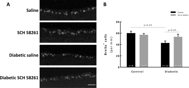
Figure 6.
A2AR antagonist attenuates RGC loss in a mouse model of diabetic retinopathy. (A) The number of RGCs was evaluated in vertical sections by immunolabeling against Brn3a (grey). The nuclei were stained with DAPI (blue). Representative images are depicted. (B) The number of Brn3a+ cells were counted, and the results were normalized to the length of the section; from 7–13 animals. One-way ANOVA test, followed by Holm-Šídák multiple comparison test. GCL: ganglion cell layer; INL: inner nuclear layer; ONL: outer nuclear layer. Scale bar: 100 µm.

