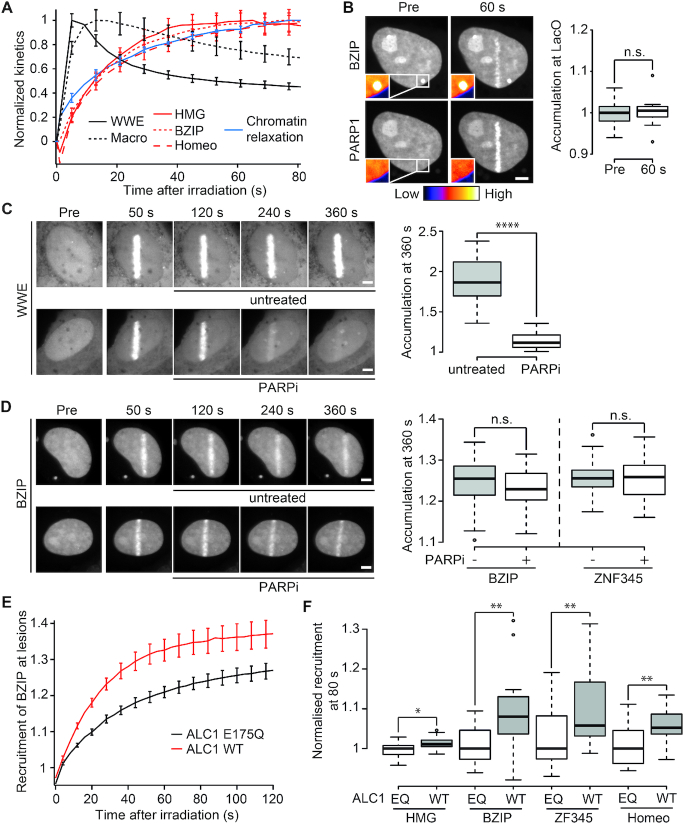
Figure 3.
Recruitment of DNA-binding domains to sites of DNA damage is not triggered by PAR-binding. (A) Normalized recruitment kinetics of PAR binding domains GFP-WWE and YFP-macroH2A1.1 macrodomain (black) and DNA-binding domains GFP-BZIP, GFP-HMG and GFP-Homeo (red) in addition to normalized chromatin relaxation kinetics (blue). (B) GFP-BZIP tethered to the LacO array is unable to recruit PARylated mCherry-PARP1 after DNA damage induction. Insets, pseudocolored according to the look-up table displayed below panel B, show magnification of the LacO array. Boxplots show mCherry-PARP1 intensity at the LacO array quantified pre- and 60 s post-damage. The average intensity at the spot is corrected for background and normalized to average intensity of the nucleus. (C, D) Confocal images showing accumulation of GFP-WWE (C) or GFP-BZIP (D) at sites of DNA damage. Cells were left untreated or treated with PARPi 60 s after DNA damage induction. Boxplots show levels of GFP-WWE, GFP-BZIP or ZF345 accumulation at sites of damage 360 s after microirradiation. (E) Recruitment of the GFP-BZIP to sites of DNA damage induced by laser microirradiation in cells overexpressing WT ALC1 (red) or ATPase dead ALC1 (E175Q, black). (F) Recruitment intensity of GFP-tagged DNA-binding domains at sites of DNA damage in cells overexpressing WT ALC1 or ATPase dead ALC1 (E175Q) measured 80 s after DNA damage induction. For each domain, the recruitment has been normalized to the median recruitment in the E175Q expressing cells. Scale bars = 4 μm

