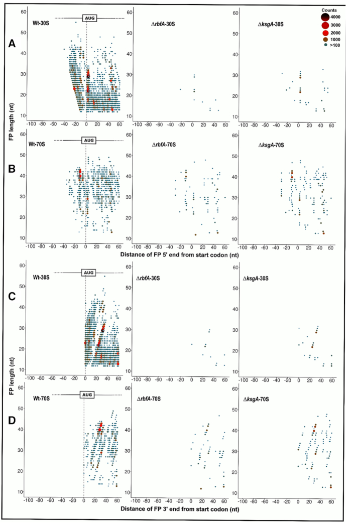
Figure 4.

Metagene analysis for translation initiation complex. (A–D) 5′ ends of the FPs derived from 30S (A) and 70S (B) bound mRNA as well as the 3′ ends of the FPs derived from the 30S (C) and 70S (D) from Wt, ΔrbfA and, ΔksgA, respectively, are mapped against the known start codon of the coding sequences in E. coli. The first base of the start codon is aligned to the ribosome P site. The size and colour of the points correspond to the frequency of occurrence of FPs. Only those FPs with at least 100 reads are represented here.
