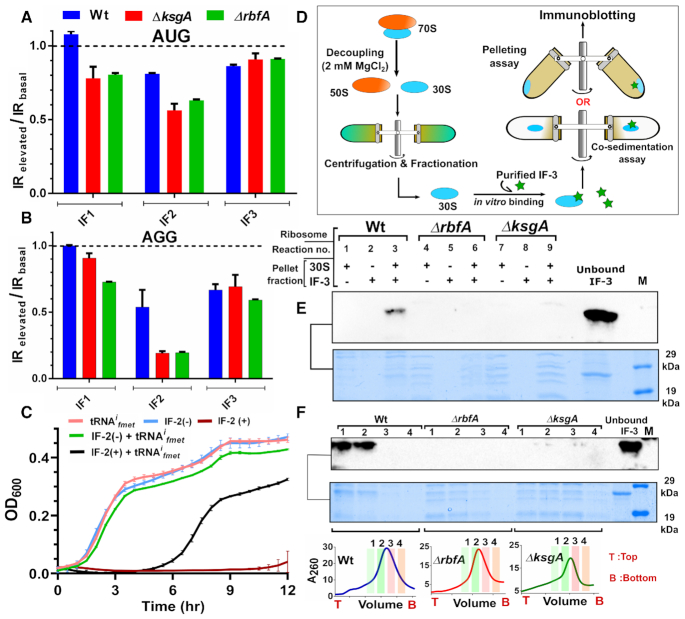
Figure 7.
Premature ribosomes evade translation quality control by initiation factors. (A and B) Bar plots representing the ratio of translation initiation rates (IRs) in cells harbouring elevated levels of IFs (IRelevated) to translation initiation rates in cells with basal levels of IFs (IRbasal) from AUG start codon (A) and AGG start codon (B). A ratio of the two translation initiation rates, derived from three independent time-course experiments are presented here. The curve fittings used to derive the respective IRs are presented in the Supplementary Figure S6. (C) Growth comparison for Wt transformed with vectors carrying genes encoding IF2 and tRNAifmet. IF2 production was induced at time = 0 by adding Atc (vide Methods) and this is indicated as IF2(+), whereas cells grown without the addition of Atc are represented as IF2(-). Overproduction of tRNAifmet was constitutive. (D) An outline of the strategy used to study the binding of IF3 to 30S subunits from Wt, ΔrbfA and ΔksgA. 30S subunits engaged in translation were purified by cycles of ultracentrifugation and IF3 binding to 30S subunits was tested using co-migration or pelleting assays. (E) Immunoblot for the pelleting assay using strep-tagged IF3 and 30S particles from Wt, ΔrbfA and ΔksgA is shown. Reaction mixtures containing 30S and IF3 either separately or in combination were ultracentrifuged on sucrose cushions. Pellet fractions from the respective reactions were resolved using SDS-PAGE and the indicated area was probed using an anti-strep antibody. The reaction components are indicated on top of the gel with the respective source of the ribosomes. Two picomoles of IF3 was directly loaded in the lane marked as ‘Unbound IF3’ as a control. Protein markers are loaded in the lane ‘M’ and the respective molecular weights are shown on the right. (F) Immunoblot for the co-migration assay using strep-tagged IF3 and 30S particles from Wt, ΔrbfA and ΔksgA is shown. A reaction mixture comprising 30S and IF3 was loaded on a 10–50% sucrose gradient, which was followed by ultracentrifugation and fractionation. Four ribosomal fractions were TCA precipitated and resolved using SDS-PAGE and the indicated area was developed using immunoblotting. The reaction components and the respective fractions are indicated above the blot and the respective ribosomal profiles and the fractions are indicated below the gel. Two picomoles of IF3 was loaded in the lanes indicated as ‘Unbound IF3’. A protein size standard containing strep tagged proteins is loaded in the lane ‘M’ and the corresponding molecular weights are indicated on the right.

