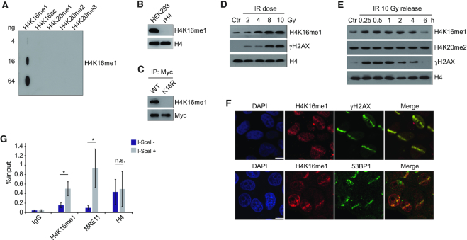
Figure 1.
H4K16me1 levels increase in response to DNA damage. (A) Slot blot assay showing high specificity of the H4K16me1 antibody. Cross reactivity with peptides carrying other modifications was not detected. (B) H4K16me1 expression in HEK293 cells. Recombinant Histone H4 (rH4) was used as a negative control. (C) HCT116 cells were transfected with a Myc-tagged H4 WT or K16R mutant vector for 3 days before anti-Myc tag immunoprecipitation and western blotting. (D) Western blot analysis of histones extracted from HCT116 cells 1 h after exposure to increasing doses of X-ray irradiation (IR). Ctr, no IR. (E) Western blot analysis of histones extracted from HCT116 cells after X-ray irradiation at 10 Gy and re-cultured for the indicated time. (F) HeLa cells were subjected to micro-irradiation, then fixed 10 min later and immunostained with the indicated antibodies. Scale bars: 10 μm. (G) DR-GFP U2OS cells were transiently transfected with I-SceI and subjected to ChIP assay 24 h later with the indicated antibodies. The IP/input% from three independent experiments is shown as the means ± SD. n.s., not significant, *P < 0.05 (Student's t-test).

