Figure 1.

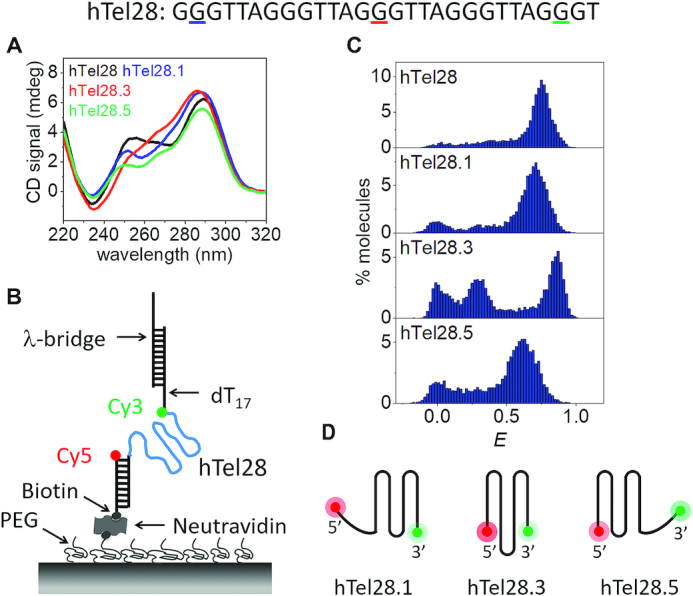
Conformational analysis of a five-repeat telomeric DNA (hTel28). (A) CD spectra of hTel28, hTel28.1, hTel28.3 and hTel28.5 in 100 mM K+. The G to T mutation sites in hTel28 corresponding to hTel28.1, hTel28.3 and hTel28.5 are underlined in colors matching the figure legend. (B) Schematic diagram of hTel28 construct for smFRET. The 5′ extension to the 28 nt long human telomeric repeat GGG(TTAGGG)4T was annealed to a 18 nt long biotinylated strand and immobilized on a PEG-passivated quartz surface through biotin-neutravidin interaction. The 5′ end of the biotinylated strand is labeled with Cy5 (acceptor). FRET was measured between Cy3 and Cy5. The main hTel28 strand is labeled with Cy3 (donor) at the 3′ end of the telomeric repeat and is followed by dT17 and an 18 nt extension that is annealed to 30 nt long λ-bridge. (C) E histograms of hTel28, hTel28.1, hTel28.3 and hTel28.5 in 100 mM K+ concentration. (D) Schematic representation of GQ formation after site-specific G to T mutation of hTel28.
