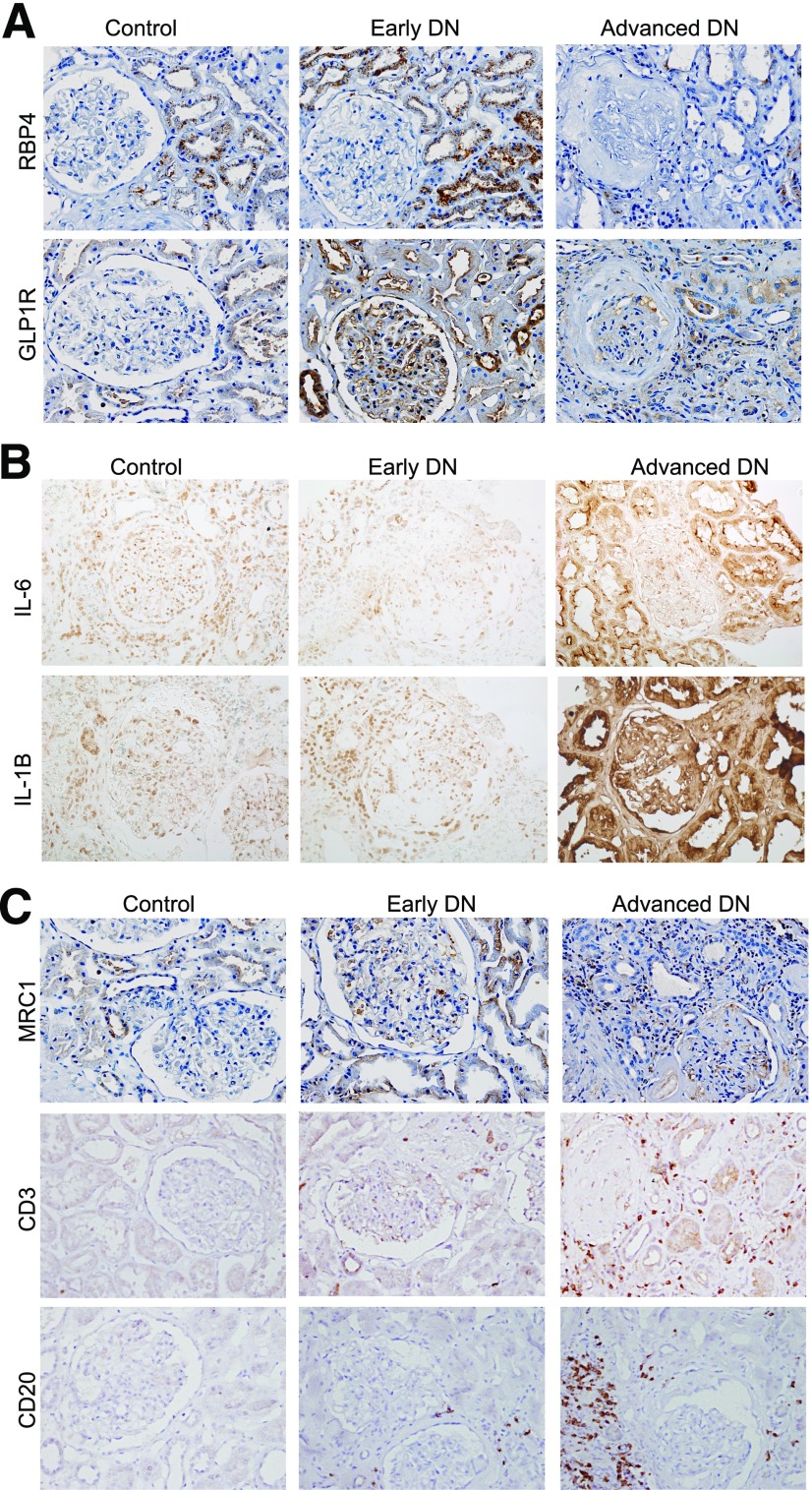
Figure 8.
Immunostaining of genes that are altered in DN. A: Representative images of RBP4 and GLP1R immunostaining in control and DN kidneys. B: Representative images of IL-6 and IL-1B immunostaining in control and DN kidneys. C: Representative images of CD20, CD3, and MRC1 immunostaining in control and DN kidneys. Semiquantification of immunostaining is shown in Supplementary Fig. 8.

