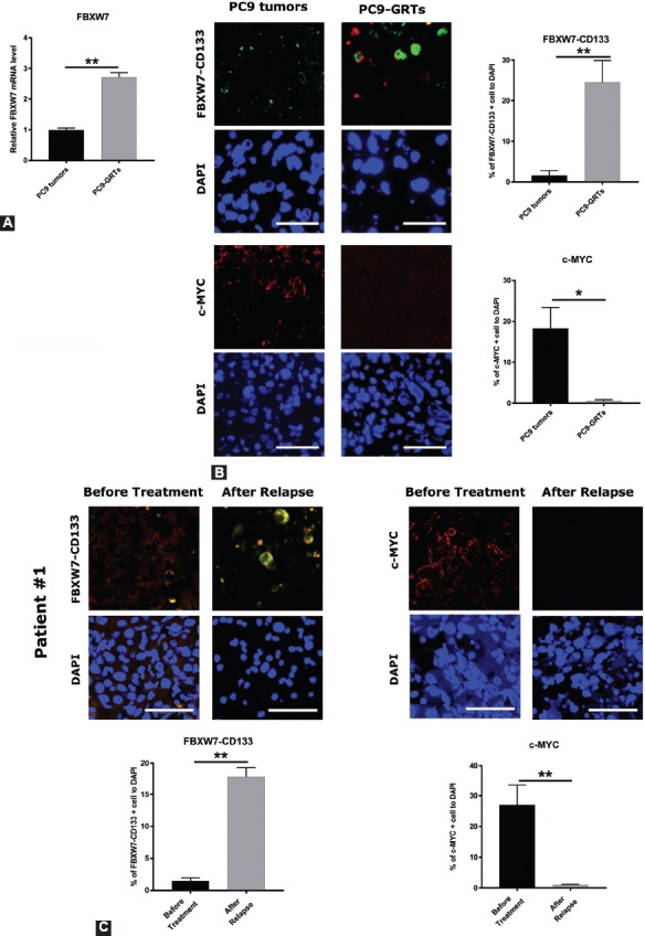
FIGURE 4.

The expression of F-box/WD repeat-containing protein 7 (FBXW7) was higher and that of c-MYC was lower in CD133-positive cells in gefitinib-resistant tumors (GRT) of PC9 in vivo as well as in lung cancer specimens from NSCLC patients who had acquired resistance to gefitinib. (A) PC9 cells were injected into NOD/Shi-scid/IL-2Rcnull (NOG) mice subcutaneously followed by intraperitoneal injection (6 times/week) of 20 mg/kg of gefitinib or vehicle after the tumor volume reached 75 mm3. After 14 to 17 days of gefitinib treatment, the tumors still remained (the mean size of the tumor was 35.3 mm3). Tumors were taken, and quantitative reverse transcription polymerase chain reaction (qRT-PCR) was carried out with FBXW7 primers, in PC9 tumors and PC9-GRT. Data were normalized to actin expression and represent mean ± SD. **p <0.01. (B, left) The expression levels of FBXW7 in CD133-positive cells and c-MYC in PC9 tumors and PC9-GRTs were examined by immunohistochemistry. (B, right) The ratio of FBXW7- and CD133-positive cells and c-MYC-positive cells. Cell nuclei were stained with DAPI (blue). Images were obtained using a ZEISS LSM 780 system. Five pictures were taken from each group and the number of cells expressing FBXW7 and c-MYC in CD133-positive cells was counted *p < 0.05, **p < 0.01. Scale bar indicates 25 µm. (C, top) The expression of FBXW7 in CD133-positive cells and c-MYC in pairs of tumor specimens prior gefitinib treatment and specimens with acquired resistance to gefitinib from NSCLC patients was examined by immunohistochemistry. (C, bottom) The ratio of FBXW7 in CD133-positive cells and c-MYC-positive cells. Cell nuclei were stained with DAPI (blue). Images were obtained using a ZEISS LSM 780 system. Five pictures were taken from each group, and the number of CD133-positive cells that expressed FBXW7 and the number of cells that expressed c-MYC was counted. *p < 0.05, **p < 0.01. Scale bar indicates 25 µm.
