Figure 2.
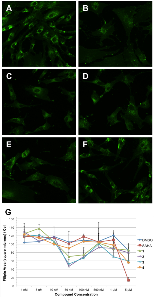
Comparison of GEX1A analogues in NPC1 mutant fibroblasts. GM18453 cells were incubated with each compound for 48 hours and subsequently fixed, stained with filipin (green false color), and imaged by fluorescence microscopy. A) DMSO; B) SAHA, 5 μM; C) GEX1A 1, 50 nM; D) GEX1A methyl ester 2, 50 nM; E) C18-OAc GEX1A methyl ester 3, 50 nM; F) C18-oxo GEX1A methyl ester 4, 50 nM. G) Dose dependence of cholesterol clearance activity. Filipin area sum and cell count were measured in fluorescence microscopy images following treatment with the indicated compound (or vehicle control) for 48 hours. Data shown are derived from four independent replicates, and four images were acquired for each condition in each experiment. Error bars represent standard deviation between images.

