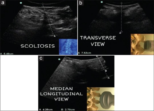
Figure 5.

Case 1 (a): Scoliosis, the posterior complex is deviated from midline depicting rotated vertebrae in ultrasound transverse view Case 2,3 (b,c): Variability in epidural depth irrespective of obesity. Use of ultrasound in measuring epidural depth in both transverse and median longitudinal view and preventing accidental dural puncture in case 3 (c) as epidural depth was only 2.7 cm
