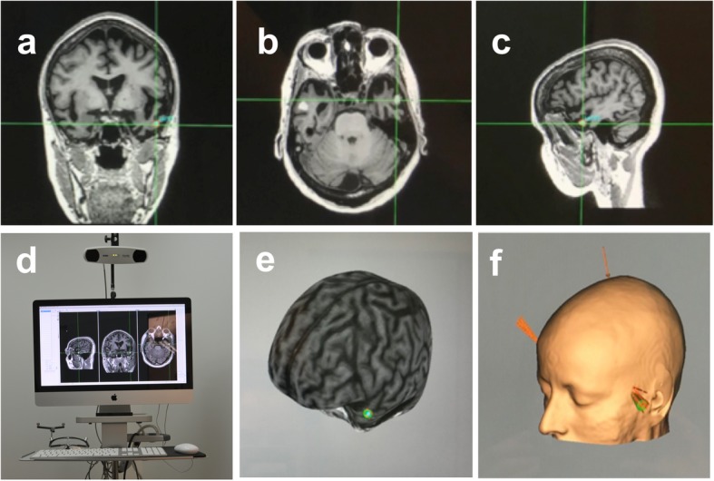
Fig. 2.

The three upper panels show, respectively, a coronal, b axial and c sagittal sections from magnetic resonance imaging (MRI) in a representative patient with semantic dementia (SD). The crosshair signals the location of the stimulation target in the left Anterior Temporal Lobe (ATL). d MRI-based frameless stereotactic navigation system device (Brainsight) employed for accurate targeting of the left ATL in SD patients. 3D brain curvilinear e cortical and f skin reconstructions from the MRI of a patient, provided by the neuronavigation system with a sphere indicating the left ATL target aimed by the stimulation
