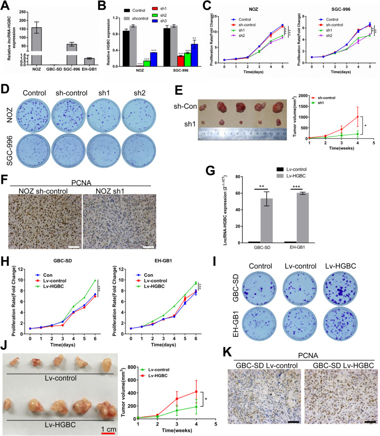
Fig. 2.
LncRNA-HGBC promotes GBC cell proliferation and tumor growth. a The relative lncRNA-HGBC expression was measured using qRT-PCR in the indicated GBC cell lines. b LncRNA-HGBC expression level was detected in NOZ and SGC-996 cells by qRT–PCR after viral infection. *P < 0.05. c Cell proliferation assays for NOZ and SGC-996 cells expressing shRNA sh1, sh2 or the negative control (sh-control) were determined using CCK-8 assays. d Typical photographs of colony formation assays of lncRNA-HGBC knockdown or control control NOZ (top) and SGC-996 cells (bottom) were shown. e Effects of lncRNA-HGBC knockdown in NOZ cells on tumor growth in vivo. Left, representative images of tumors formed in nude mice (n = 5). Right, tumor volumes were measured once a week and tumor growth curves are summarized in the line chart (*P < 0.05). f PCNA expression was examined in sections of NOZ xenografts by immunohistochemistry. Scale bars, 100 μm. g lncRNA-HGBC expression level was determined in GBC-SD and EH-GB1 cells by qRT–PCR. h Cell proliferation assays for GBC-SD and EH-GB1 cells expressing lncRNA-HGBC or the control. i Colony formation assays of lncRNA-HGBC-overexpressing or control GBC-SD (top) and EH-GB1 cells (bottom). j Images of tumor formation in nude mice (n = 5) injected subcutaneously with GBC-SD cells overexpressing lncRNA-HGBC (bottom) or the control (top). Tumor volumes were measured once a week and tumor growth curves are summarized in the line chart.(*P < 0.05). k PCNA expression in sections of GBC xenografts was determined by immunohistochemistry. Scale bars, 100 μm

