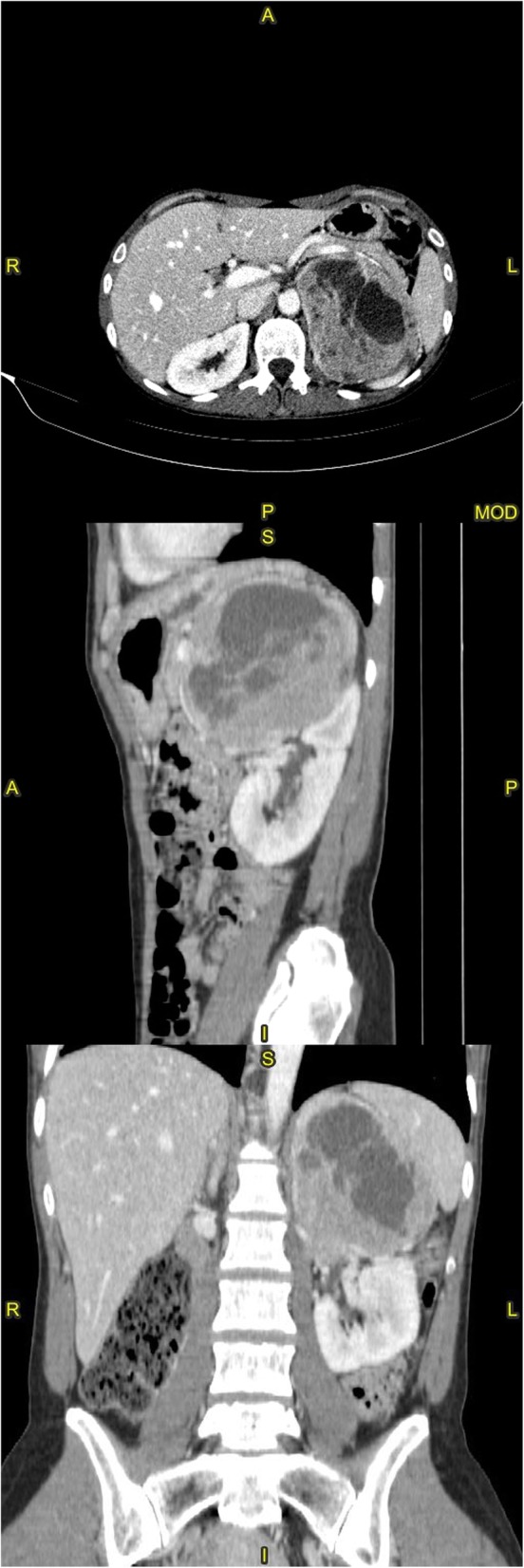
Fig. 1.

Multiplanar reconstruction of a contrast-enhanced abdominal computed tomography scan. The left adrenal mass has a maximum diameter of 11.2 cm, and is seen displacing the gastric fundus antero-superiorly and the spleen laterally; cleavage from the kidney is unclear around the pelvis in the coronal image, suggesting cancerous infiltration
