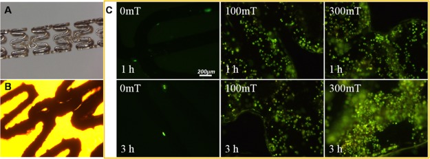
Figure 8.
(A) Stereo-micrograph of the iron stent. (B) Bright-field image of the iron stent; (C) EPC adhesion onto iron stents assisted by Fe3O4@CA–CD34 and different EMF (0, 100 and 300 mT) for 1 and 3 h under flow conditions in vitro; cellular labeling was visualized histochemically with rhodamine 123 staining.

