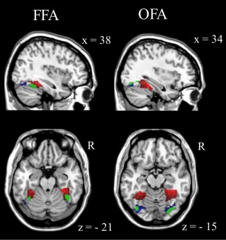
Figure 3.

Overlays of the task‐based functionally defined face sensitive areas (green; according to Fig. 2) with the resting‐state derived polarity distributions (according to Fig. 1a) of bilateral amygdala rs‐FC (entire sample) revealed that positive rs‐FC (red) in the anterior/medial FFG, respectively, corresponded to the fusiform face area and that negative rs‐FC (blue) in the posterior FFG, respectively, corresponded to the occipital face area (Table 4). [Color figure can be viewed in the online issue, which is available at http://wileyonlinelibrary.com.]
