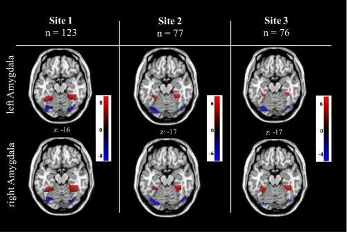
Figure 4.

Analyses in the independent scanner sites confirmed reliability as revealed by similar patterns of amygdala rs‐FC polarity distribution within the FFG in all three sites, whereas in site three the right sided cluster based on negative right amygdala rs‐FC in the posterior FFG did not survive (P = 0.061) the cluster correction with P < 0.05 (FWE, small‐volume). [Color figure can be viewed in the online issue, which is available at http://wileyonlinelibrary.com.]
