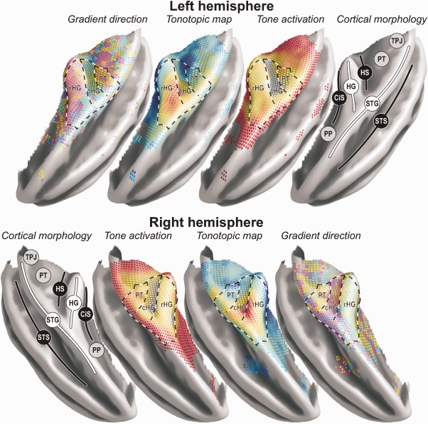
Figure 7.

A three‐dimensional rendering of key results on the group‐average semi‐inflated temporal lobe surface. Results were copied from the surface‐based analysis with 5‐mm smoothing, employing colour‐codes identical to those shown in Figure 4a,c,d. Notable morphological features include the planum polare (PP), circular sulcus (CiS), Heschl's gyrus (HG), Heschl's sulcus (HS), planum temporale (PT), temporoparietal junction (TPJ), superior temporal gyrus (STG), and superior temporal sulcus (STS). The superimposed dashed lines delineate the approximate outline of two fields on rostral (rHG) and caudal (cHG) HG that could be clearly distinguished on the basis of the tonotopic organisation. Evidence for at least one additional field labelled PT was additionally found further posteriorly. On the lateral side, adjacent to STS, the organisation was ambiguous (DISCUSSION section).
