
Figure 1.
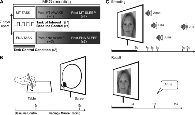
(A) MEG signal was acquired with 151 axial gradiometers while participants performed either the MT task or the task control condition, a FNA task and were subsequently asked to sleep while remaining in supine position for 80 min. Analyses were performed over the duration of the tasks, over the first 15 min of wakefulness and over the first 15 min after the onset of NREM Stage 2 sleep. In a counter‐balanced order, the procedure was repeated for the other task after 7 days. The numbers in italics indicate the periods that were analyzed: t1 was the MT task, r2 was the resting‐state baseline of the MT task, t2 was the FNA task, w1 and w2 were the periods of wakefulness following the MT task and the FNA task, respectively, and s1 and s2 were the sleep periods following the MT task and the FNA task respectively. (B) The task of interest was the MT task: Participants had to trace the outline of a projected circle as fast and accurately as possible by moving a pen on a graphics tablet to control the position of a projected dot. In half of the trials, the location of the dot was mirrored about the y‐axis. Participants were asked to rest during 5 s baseline periods alternating with tracing. This period was subsequently used to test that the activation was specific to task execution and was not present during the baseline resting‐state control condition. (C) The task control condition was the FNA task: During the encoding phase, participants observed 34 faces, each presented for 5 s. Two seconds after the disappearance of each face, three possible names and finally the number (1, 2, or 3) of the name to be remembered were given. During the recall phase, participants had to name each presented face.