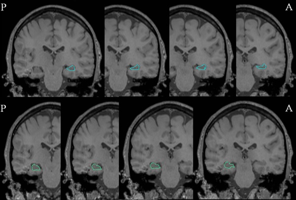
Figure 1.

Radial mapping analysis: hippocampal segmentation. Coronal magnetic resonance imaging (MRI) slices from a healthy control in posterior‐anterior direction. Representative manual tracings of the right (light‐blue, top row) and left (light green, bottom row) hippocampus are shown.
