
Figure 1.
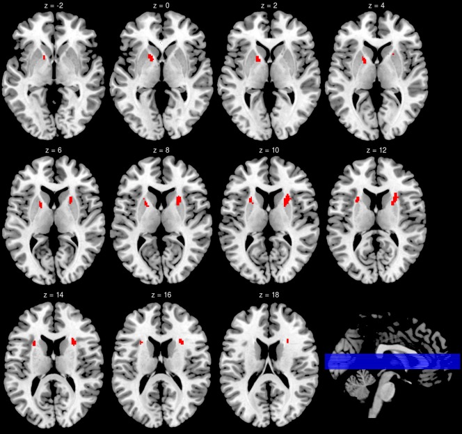
Brain areas that showed reduced (right hemisphere) or a trend for reduced (left hemisphere) FA values for insomnia patients. For the illustration of the cluster in the left hemisphere, the minimal cluster size for this figure was set at 320 mm³ (40 voxels) at a voxel threshold of P < 0.005 (T > 2.93).