
Figure 1.
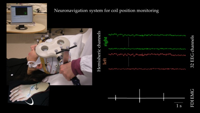
Experimental setup. Left neuronavigated TMS setup: the subject lies relaxed on a semireclined chair and the coil position is monitored throughout the session duration. The hand with surface electrodes for FDI muscle recordings is evidenced in the inset (white arch, ground symbol indicates belly‐tendon montage, while on the wrist dorsum is the ground electrode). Right EEG and EMG signals after artifact removal. [Color figure can be viewed in the online issue, which is available at http://wileyonlinelibrary.com.]