
Figure 1.
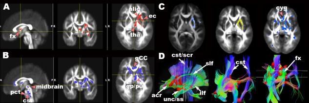
Maps of significant group differences of voxel‐wise analyses of FA (A (FWE=5%, TFCE P ≤ 0.05), B (FWE=5%, TFCE P ≤ 0.06)), trace (C left (light blue, FWE=5%, c > 2.3, P ≤ 0.05)), radial diffusivity (C middle (yellow, FWE=5%, c > 2.5, P ≤ 0.05)), and parallel diffusivity (C right (blue uncorrected P ≤ 0.05)) and reconstructed tracts (D) using lower FA (A) as seeds in axial, sagittal and coronal views overlaid on the mean FA template (1 × 1 × 1 mm3). acr, anterior region of corona radiata; alic, anterior limb of internal capsule; bCC, body of corpus callosum; cst, corticospinal tract in brainstem; cyg, cingulum; gp, globus pallidus; pu, putamen; ec, external capsule; fx, fornix; ilf, inferior longitudinal fasciculus; pct, pontine crossing tracts; scr, superior region of corona radiata; slf, superior longitudinal fasciculus; ss, sagittal stratum, including inferior longitudinal fasciculus and inferior fronto‐occiptal fasciculus; tha, thalamus; unc, uncinate fasciculus.