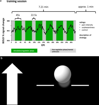
Figure 1.

a: Course of one training trial. Over the course of one training trial (290 echo planar imaging sequences) the participants attempted either upregulation or downregulation of the activation in the target area during six regulation phases each lasting 45 s (green) while receiving painful electrical stimulation. Between the regulation phases, that is, in the nonregulation phases (gray) each lasting 22.5 s, the subjects performed mental arithmetic. An exemplary blood oxygenation level‐dependent (BOLD) time course is superimposed in black. b: Exemplary feedback screen. The arrow on the left indicated the direction in which the ball should be moved. The amplitude of the ball was computed as the difference between the percent signal change in the target region of interest and an unrelated region. The ball color was either blue or yellow for the rostral anterior cingulate cortex (rACC) and the posterior insula (pInsL) conditions. The color assignment to the target regions was randomized.
