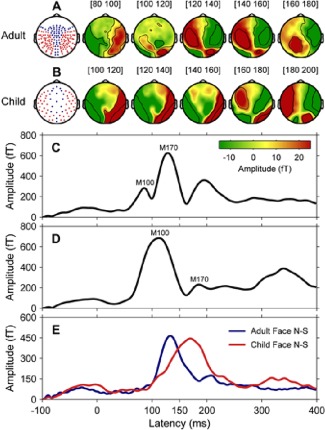
Figure 2.

Event‐related magnetic fields in adults (N = 14) and children (N = 15). (A) Schematic diagram of sensor layout for 160‐channel adult MEG system and surface topography in adults for the difference response at 20 ms stepwise from 80 to 180 ms, encompassing the temporal window of M100 (83 ms) and M170 (148 ms). (B) Schematic diagram of sensor layout for 64‐channel child MEG system and surface topography in children for the difference response at 20 ms stepwise from 100 to 200 ms, encompassing the temporal window of M100 (110 ms) and M170 (180 ms). In both MEG sensor layouts, red dots indicate the temporal–occipital regions where the M170 response was maximal. Note here the child system does not have frontal region coverage. (C) Global field power of grand mean ERFs to faces in adults. (D) Global field power of grand mean ERFs to faces in children. (E) Global field power of grand mean ERFs to faces minus ERFs to scrambled faces (Face N–S) for adults (blue line) and children (red line). [Color figure can be viewed in the online issue, which is available at http://wileyonlinelibrary.com.]
