Fig. 1.
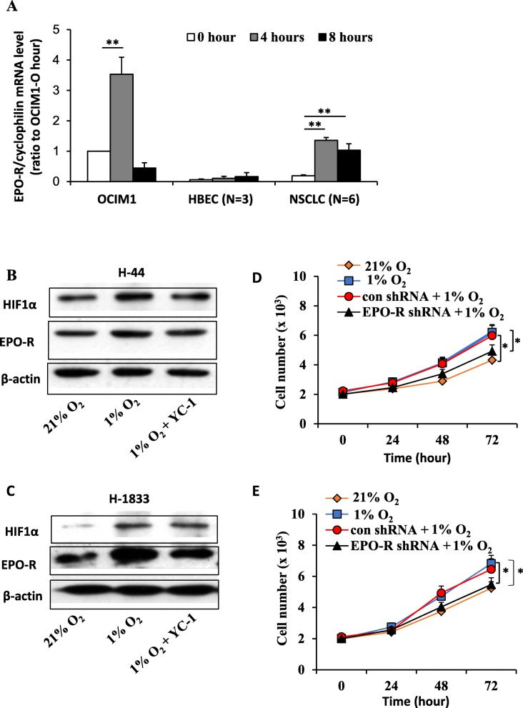
Hypoxia-induced EPO-R overexpression promotes cell proliferation in NSCLC. a Hypoxia induced EPO-R expression in NSCLC cells. Three HBEC (HBEC3KT, HBEC4KT and HBEC6KT) and 6 NSCLC (A549, H44, H2073, H1819, H1833, H3122) cell lines were treated with 1% hypoxia or room air for 0, 4 or 8 h. The erythroleukemia line OCIM-1 was used as a positive control. The mRNA level was determined by real-time RT-PCR with cyclophilin as an internal control. Mean ± SEM; ** P < 0.01. b and c Expression of EPO-R protein was upregulated under hypoxia in H44 (b) and H1833 cells (c), which was diminished by treatment with HIF1α inhibitor YC-1. Protein expression was determined by Western blots and β-actin was used as a loading control. d and e MTT assays showed that hypoxic treatment promoted H44 (d) and H1833 (e) cell growth, and knockdown of EPO-R with specific shRNA abolished these effects. The data are representative of three experiments. Mean ± SEM; *P < 0.05

