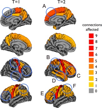
Figure 4.

Cortical regions included in the affected sub‐network at T = 1 and T = 2. The regions are colored based on the number of affected connections. The blue circles indicate the newly affected cortical regions at T = 2 compared with T = 1 (A: superior frontal right; B: postcentral and superior parietal right; C: rostral middle frontal, pars orbitalis, pars triangularis, and pars opercularis right; D: temporal pole, superior, and middle temporal right, insula right [not visible]; E: rostral middle frontal and pars triangularis left; and F: superior parietal left). [Color figure can be viewed in the online issue, which is available at http://wileyonlinelibrary.com.]
