
Figure 5.
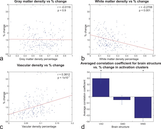
Cluster‐wise (N = 199) analysis of relationship between (A) GMD, (B) WMD, and (C) VAD and T‐fMRI % change during activation for 19 subjects. (D) Average correlation across clusters and subjects between structural density and T‐fMRI % change during activation. VAD and WMD correlations were statistically significant (P < 0.05). Error bars reflect SEM. [Color figure can be viewed in the online issue, which is available at http://wileyonlinelibrary.com.]