
Figure 3.
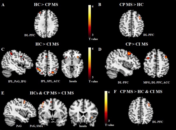
Brain regions showing significantly different fMRI activations with increasing N‐back task difficulty in cognitively impaired (CI) and cognitively preserved (CP) patients with multiple sclerosis (MS) vs. healthy controls (HC): (A) HC vs. CP MS patients; (B) CP MS patients vs. HC; (C) HC vs. CI MS patients; (D) CP vs. CI MS patients; (E) HC and CP MS vs. CI MS patients (conjunction analysis); (F) CP MS vs. CI MS patients and HC (conjunction analysis). Images are in neurological convention. Abbreviations: IPL, inferior parietal lobule; SPL, superior parietal lobule; PcG, precentral gyrus; IFG, inferior frontal gyrus; MFG, middle frontal gyrus; DL‐PFC, dorsolateral prefrontal cortex; SMA, supplementary motor area; ACC, anterior cingulate cortex.