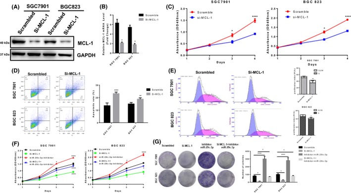
Figure 7.

Mcl‐1 expression is up‐regulated in gastric cancer tissues and promotes gastric cancer cell growth. A, The knockdown efficiency of MCL‐1 was determined by Western blotting in SGC‐7901 and BGC‐823 cells. B, MCL‐1 mRNA levels were determined by real‐time quantitative PCR in MCL‐1 knockdown SGC‐7901 and BGC‐823 cells. C, Scrambled siRNA or si‐MCL‐1 was transfected into SGC‐7901 and BGC‐823 cells, and cell proliferation ability was measured by CCK8. D, Scrambled siRNA or si‐MCL‐1 was transfected into SGC‐7901 and BGC‐823 cells and used to detect apoptosis rate by flow cytometry. E, Scrambled siRNA or si‐MCL‐1 was transfected into SGC‐7901 and BGC‐823 cells, and cell cycle was analysed by flow cytometry. F, The proliferation ability of the cells was determined by CCK8 after co‐transfection of si‐MCL‐1, miR‐29c‐3p inhibitor or scrambled siRNA in SGC‐7901 and BGC‐823 cells. G, The proliferation ability of the cells was determined by colony formation assay after co‐transfection of si‐MCL‐1, miR‐29c‐3p inhibitor or scrambled siRNA in SGC‐7901 and BGC‐823 cells. Values represent the mean ± SEM of three independent experiments. *P < .05, **P < .01, ***P < .001
