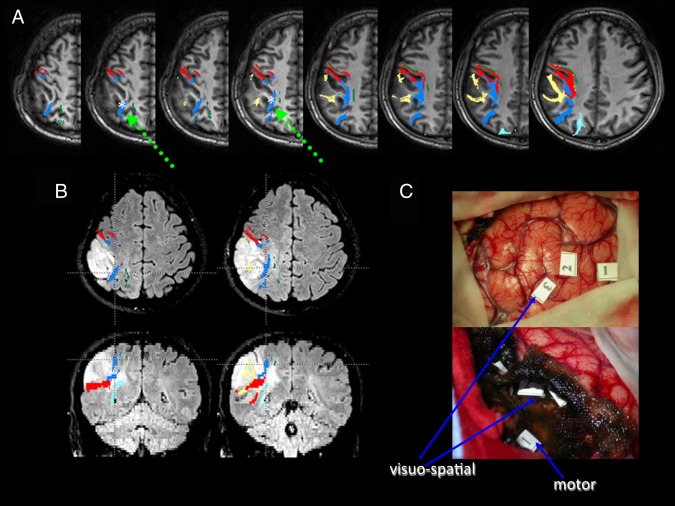
Figure 1.

Patient P4. A) Preoperative deterministic tractography reconstructions superimposed on axial T1‐weighted images; SLF I fibers are depicted in green, SLF II in blue, SLF III (that includes fibers of the horizontal part of the AF) in yellow, AF in red, and IFOF in azure. The white asterisk and the green arrow show the DES site of the SLF II, which caused a rightward bisection error during intraoperative stimulation. B) Morphologic FLAIR axial (upper) and coronal (lower) images of the tumor, showing both the displacement and the infiltration of SLF fibers along the tumor margins: specifically, the second branch was much closer to the tumor border than the third branch and the AF, which was located more laterally and ventrally, and the first branch, which was displaced superiorly. This information was available during surgery, because tractography reconstructions were loaded into the neuronavigation system. At surgery, tumor resection was immediately performed at a subcortical level close to these tracts, in order to reduce the risk of brain shift. The patient was asked to perform the bisection test, with the sites corresponding to bisection deviations being registered into the neuronavigation system. Axial and coronal sections correspond to the positive sites at stimulation. These sites were found in the subcortical white matter of the parietal lobe, in a location consistent with the trajectory of the SLF II at a dorsal level: note that fibers of this branch lie dorsally to the fibers of the AF. C) Intraoperative pictures of the surgical cavity at the beginning (upper) and at the end of tumor resection (lower); numbers correspond to the sites where DES induced intraoperatively a rightward bisection error at cortical (“3” indicates BA7b), and subcortical (SLF II) levels; “1” and “2” indicate BA 39; at a subcortical level also a motor response was induced.
