
Figure 3.
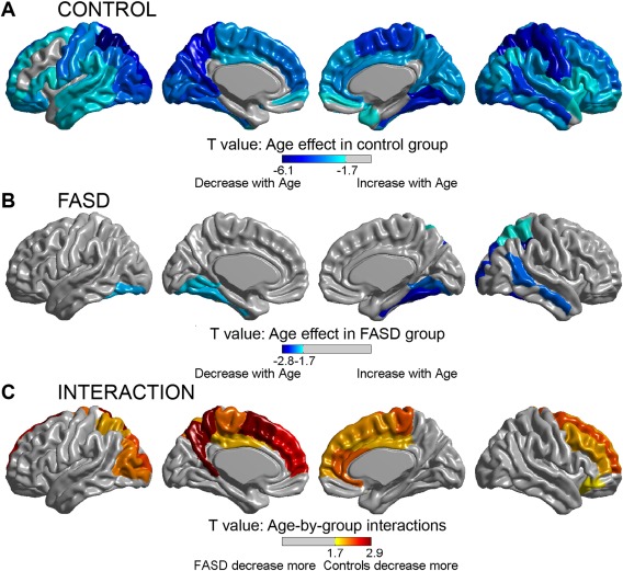
Regional age effects and age‐by‐group interactions on cortical thinning. The top two panels display AAL regions with significant effects of cortical thickness with age in the control (A) and FASD (B) groups. Significant effects of age (decreasing thickness with increasing age) are seen across most of the cortical mantle in the control group (A), but are limited to discrete temporal, parietal, and occipital regions, mostly right hemisphere, in the FASD group (B). Likewise, several regions with significant age‐by‐group interactions are found (C), indicating regional differences in the rate of change with age, with the FASD group undergoing less thinning than controls between scans. [Color figure can be viewed in the online issue, which is available at http://wileyonlinelibrary.com.]