
Figure 1.
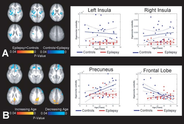
Differences in whole brain connectome hubs in children with epilepsy compared to controls. (A) Group effects show that insulae of children with epilepsy are weaker regional hubs compared to controls. (B) Age effects show that the PCC and frontal lobe demonstrate age‐related interactions in children with epilepsy. The PCC becomes more central as a function of age in controls compared with children with epilepsy, while the centrality of the frontal lobe fails to weaken with age in children with epilepsy. Axes scaled to show variance in data; steepest increases with age identified in the PCC (slopecontrols = 1.1 × 10⁻³; slopeepilepsy = 7.0 × 10⁻⁵). [Color figure can be viewed in the online issue, which is available at http://wileyonlinelibrary.com.]