
Figure 8.
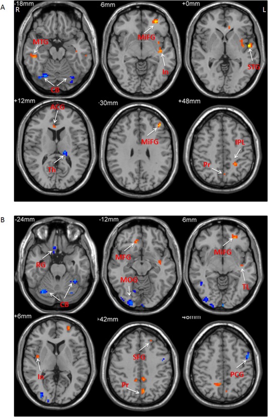
The results of correlation between ALFF and age in PD patients. The correlation between ALFF and age in the slow‐4 band (A), and slow‐5 band (B). Correlation analysis, P < 0.05, FWE corrected. Hot and cold colors indicate positive and negative correlation, respectively. L, left; R, right. ACG, anterior cingulate gyrus; CB, cerebellum; In, insula; IPL, inferior parietal lobule; MFG, medial frontal gyrus; MiFG, middle frontal gyrus; MOG, middle occipital gyrus; MTG, middle temporal gyrus; PCG, precentral gyrus; Pr, precuneus; PG, precentral gyrus; PMC, premotor cortex; RG, rectal gyrus; SFG, superior frontal gyrus; STG, superior temporal gyrus; Th, thalamus; TL, temporal lobe.