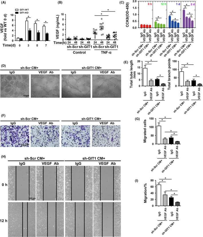
Figure 2.

GIT1 deficiency inhibits expression and secretion of VEGF in BMSCs. (A) VEGF mRNA detected by qPCR in mBMSCs by direct adherent 24 hours culture from bone marrow adjacent to the fracture site in GIT1 WT and KO mice between 0 and 7 days post‐surgery. Data are represented as means ± SEM (n = 3 for both WT and KO mice). *P < .05. (B) hBMSCs were transfected with sh‐GIT1 or sh‐Scr for 48 hours and then subjected to control sham or TNF‐α (10 ng/mL) for 24 and 48 hours. VEGF concentration was detected by ELISA in hBMSC‐CM (n = 6). *P < .05. (C) Proliferation of HUVECs cultured with hBMSCs‐CM examined by CCK8 12 hours, 1, and 2 d after TNF‐α stimulation. Data are represented as means ± SEM (n = 8). *P < .05. (D, F, H) Representative images of Matrigel tube formation, transwell and scratch wound assays with hBMSCs‐CM cultures after TNF‐α stimulation for 48 hours. Scale bar, 100 μm. (E, G, I) Quantitative analysis of tube length and branch points during tube formation (D), the number of migrated cell (F) and migration rate of HUVECs (I). Data are represented as means ± SEM (n = 6). *P < .05
