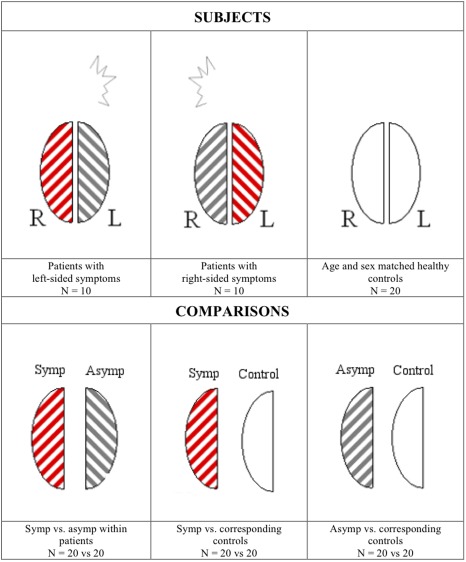
Figure 1.

General principle of the hemisphere comparison. Hemispheres of patients with visual aura symptoms consistently originating from either the right (N = 10) or the left (N = 10) hemisphere are examined. Symptomatic (symp) hemispheres (red stripes) of the patients (N = 20) are compared to their contralateral asymptomatic (asymp) hemispheres (gray stripes, N = 20). Subsequently, the symptomatic and asymptomatic patient hemispheres are compared to hemispheres of matched healthy control subjects (white). Left patient hemispheres are compared to left control hemispheres and vice versa. [Color figure can be viewed in the online issue, which is available at http://wileyonlinelibrary.com.]
