
Figure 5.
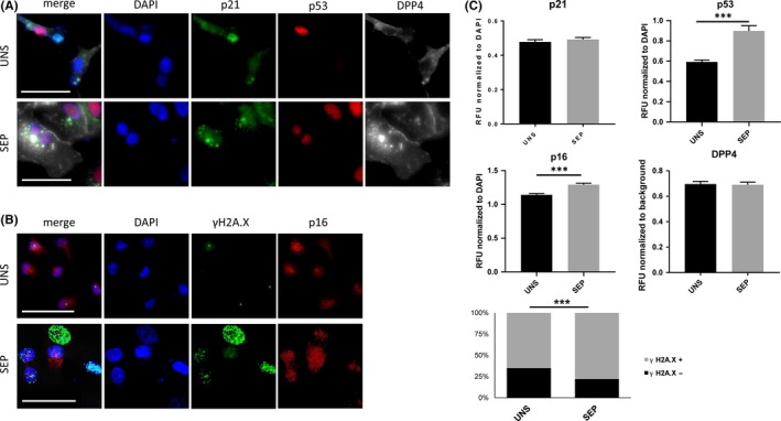
Immunofluorescent detection of selected markers of senescence. Huh‐7 cells were treated with 100 nmol/L DXR for 48 h, followed by 5 d washout. DXR‐treatment was followed by iodixanol density gradient‐based centrifugation (separated, SEP) or not (unseparated, UNS) and fixed 24 h after re‐plating. (A) Representative micrographs displaying DAPI, p21, p53 and DPP4 staining, and merge, in DXR‐treated, SEP or UNS by gradient‐based centrifugation. Scale bar represents 50 μm. (B) Representative micrographs displaying DAPI, γH2A.X and p16 staining, and merge, in control and DXR‐treated, SEP or UNS by gradient‐based centrifugation. Scale bar represents 50 μm. (C) Normalized quantification of p21, p53, p16 and DPP4 staining intensity and scoring of γH2A.X nuclei, calculated over a total of ~ 600 cells per condition. n = 4. ***P < .001