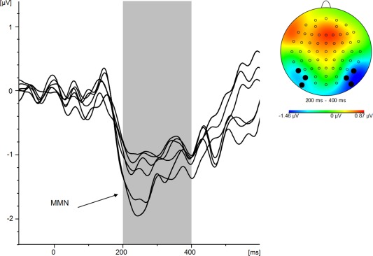
Figure 2.

Overlay of the difference potential collapsed across deviants minus neutral standard that indicates an average negativity between 200 and 400 ms. First, a butterfly plot was constructed. Next, corresponding topographical maps revealed confined negative potentials of the involved ERP components over ventral occipital cortical areas which led to the inclusion of electrodes P7, P07, P09 (left hemisphere) and P8, P08, P010 (right hemisphere). Electrodes not selected for further analysis were then removed from the butterfly plot, thus leaving included difference waveforms. [Color figure can be viewed in the online issue, which is available at http://wileyonlinelibrary.com.]
