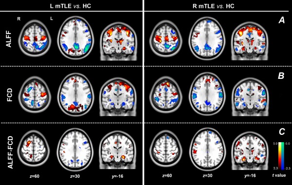
Figure 1.

Comparing results of imaging parameters between mTLE patients and HCs. Group comparison of ALFF (A) and FCD (B). In the patients with mTLE, opposite alterations of ALFF (increase) and FCD (decrease) were found in the mesial temporal lobes and insula; consistent decreases of ALFF and FCD was found in the regions of the default‐mode network; and consistent increases was found in the precentral cortices. Moreover, increased ALFF was also found in the thalamus. (C) Group comparison of ALFF‐FCD contrast map showed more pronounced group differences in the mesial temporal lobes. [Color figure can be viewed in the online issue, which is available at http://wileyonlinelibrary.com.]
