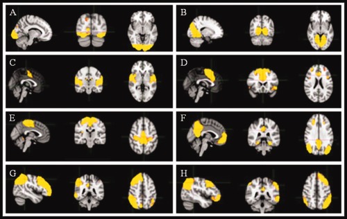
Figure 1.

Sagittal, coronal and axial slices for the main RSNs detected, overlaid onto a standard EPI functional template. (A) lateral visual, (B) medial visual, (C) auditory, (D) cognitive control, (E) sensory‐motor, (F) DMN, (G) left fronto‐parietal, (H) right fronto‐parietal.
