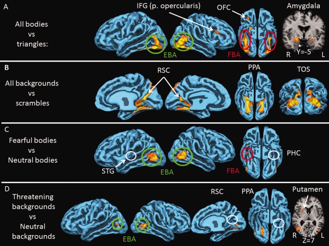
Figure 2.

Neural activity for perceiving bodies and backgrounds. A: Brain areas responding more to bodies than to triangles (P < 10−4): FBA (red); EBA (green), inferior frontal gyrus (IFG), pars opercularis, orbitofrontal cortex (OFC), and amygdala. B: Brain areas responding more to backgrounds than to scrambled images (P < 10−4): transverse occipital sulcus (TOS), parahippocampal place area (PPA), and retrosplenial cortex (RSC). C: brain areas responding more to fearful bodies than neutral bodies (P < 10−3): EBA and FBA. The superior temporal gyrus (STG) and parahippocampal cortex (PHC) responded more to neutral than to fearful bodies. D: Brain areas responding more to threatening backgrounds than to neutral backgrounds (P < 10−3): EBA right posterior PPA, and putamen. The right RSC and left anterior PPA responded more to neutral than to threatening backgrounds. Cortical activity is shown on a reconstruction of the average cortically aligned brains of all 20 participants. Gyri are shown in light blue and sulci in dark blue. Negative activation is circled in white. Coordinates refer to TAL space.
