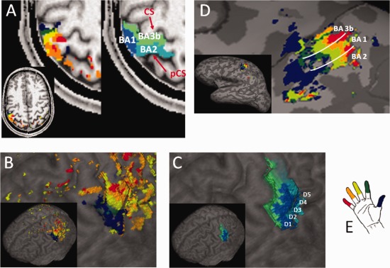
Figure 1.

Results of the finger mapping procedure. The finger maps and BAs of one representative subject are displayed (A) on a selected axial plane, (B) and (C) on the cortical surface, and (D) on the inflated brain (including a schematic representation of the borders between the three BAs). The color code for the fingers is represented in (E), while different tones of blue indicate the three functional subregions. As anatomical references, the central sulcus (CS) and postcentral sulcus (pCS) are indicated on the axial slice.
