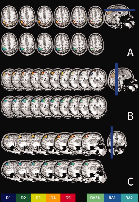
Figure 2.

A set of axial (A), coronal (B), and sagittal (C) slices extracted from the same subject as in Fig. 1, showing the representations of the five fingers and BAs. All slices are displayed relative to the scanner coordinates, and have not been reoriented relative to the bicommissural plane.
