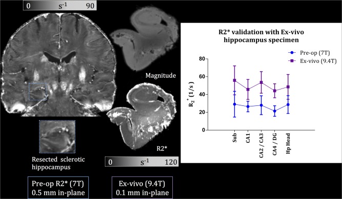
Figure 3.

Validation of pre‐operative with 9.4 T ex vivo imaging of a resected hippocampus. Left: coronal slice of the pre‐op at 7 T with a zoomed‐in view of the sclerotic hippocampus before epilepsy surgery excision. Middle: magnitude image of the patient's resected hippocampus imaged at 9.4 T resulting in a 0.1 mm isotropic resolution (top); image of the hippocampus, where the subfield delineation was performed (bottom). Right: graph of comparison between pre‐op (blue) and ex vivo (purple) values within the subfields. [Color figure can be viewed in the online issue, which is available at http://wileyonlinelibrary.com.]
