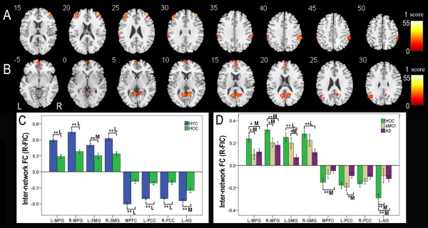
Figure 7.

Internetwork FC differences of the right FIC across groups. Voxel‐based analysis shows brain regions of the CEN (A) and DMN (B) that have internetwork FC differences (P < 0.05, FWE corrected) with the right FIC across the four groups. Each ROI with the most statistical significance in the voxel‐based analyses were then extracted. ROI analysis shows brain regions of the CEN and DMN between the HYC and HOC (C) and among the HOC, aMCI, and AD (D) that have internetwork FC differences with the right FIC across the four groups. The x‐axis represents the ROIs, and the y‐axis represents the internetwork FC of each ROI. Error bars indicate the standard error of the mean. *P < 0.05, uncorrected; **P < 0.05, Bonferroni corrected. “S,” “M,” and “L” represent small, medium, and large effect sizes, respectively. Abbreviations: AD, Alzheimer's disease; AG, angular gyrus; aMCI, amnestic mild cognitive impairment; CEN, central executive network; dACC, dorsal anterior cingulate cortex; DLPFC, dorsolateral prefrontal cortex; DMN, default‐mode network; FC, functional connectivity; FIC, frontoinsular cortex; FWE, family‐wise error; HOC, healthy older control; HYC, healthy younger control; IFG, inferior frontal gyrus; L, left; MFG, middle frontal gyrus; MPFC, medial prefrontal cortex; PCC, posterior cingulate cortex; R, right; ROI, region of interest; SMG, supramarginal gyrus; SN, salience network. [Color figure can be viewed in the online issue, which is available at http://wileyonlinelibrary.com.]
