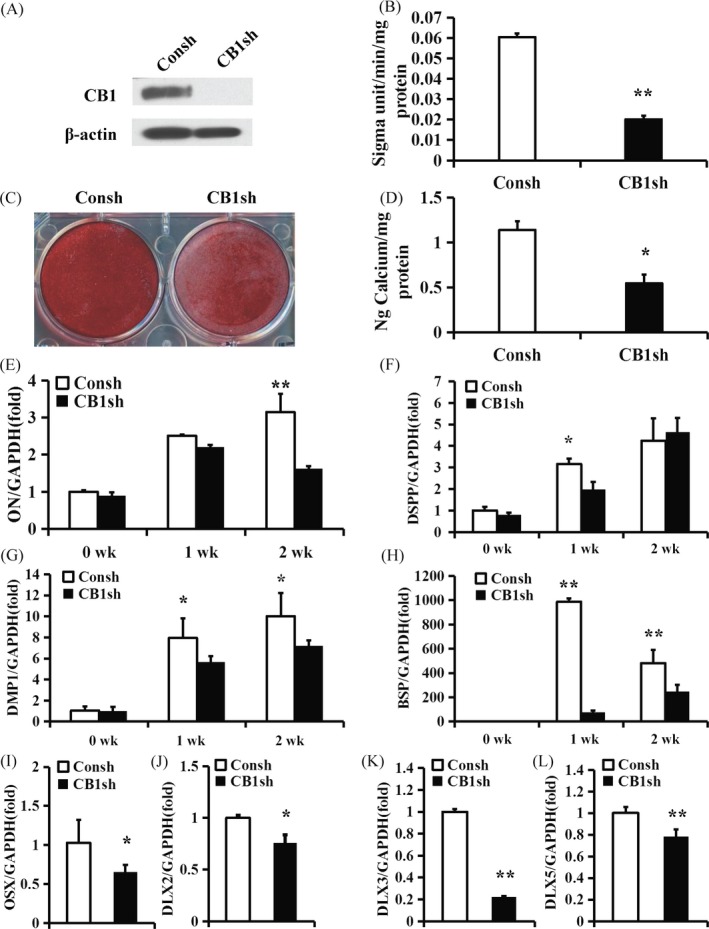
Figure 1.

CB1 knock‐down inhibited the osteo/dentinogenic differentiation of PDLSCs. (A) Western blot results showed the knock‐down efficiency of CB1 shRNA in PDLSCs. β‐actin was used as an internal control. (B) ALP activity assay. (C) Alizarin Red staining. (D) Calcium quantitative analysis. (E‐H) Real‐time RT‐PCR results of the ON (E), DSPP (F), DMP1 (G) and BSP (H) expression levels during PDLSC osteo/dentinogenic differentiation. (I‐L) Real‐time RT‐PCR results of OSX (I), DLX2 (J), DLX3 (K) and DLX5 (L) expression levels in PDLSCs. GAPDH was used as an internal control. Student's t test was performed to determine statistical significance. Error bars represent the SD (n = 3). *P ≤ .05; **P ≤ .01
