
Figure 4.
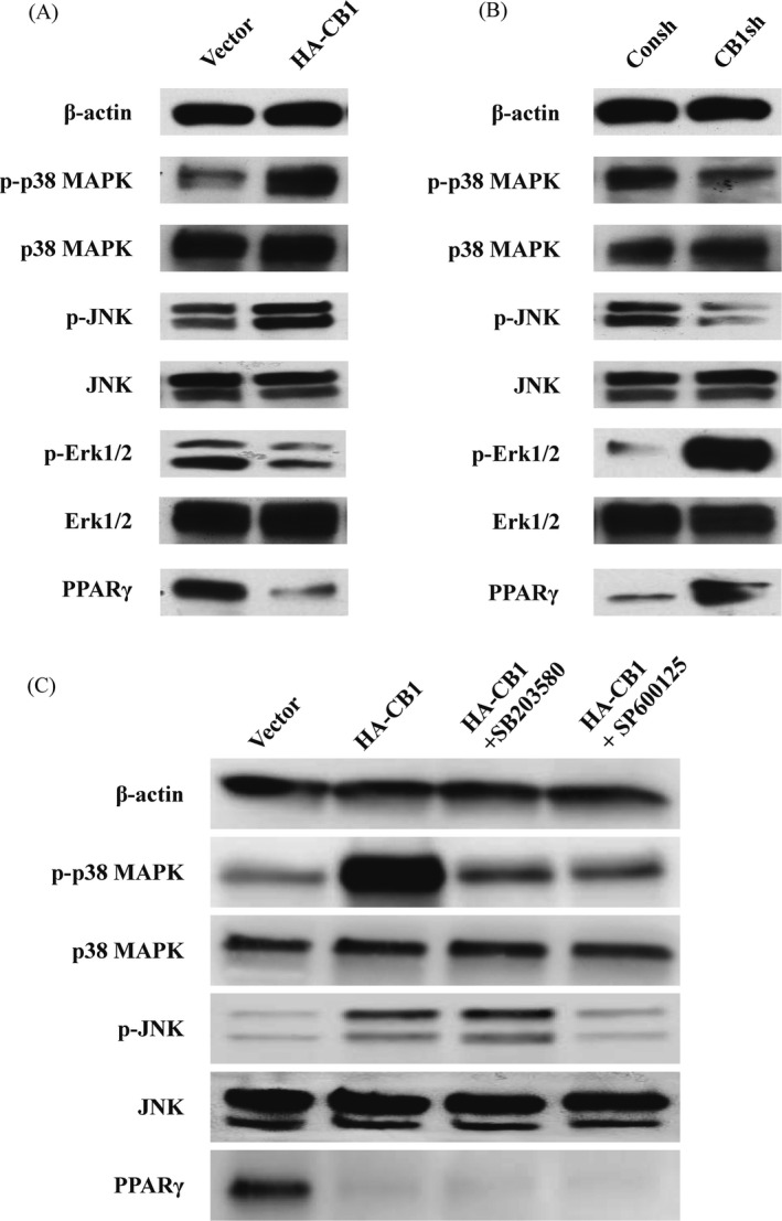
The effect of CB1 on MAPK signal pathways and PPAR‐γ in PDLSCs. A, Western blot results showed the expression of phosphorylated p38 MAPK, JNK, and Erk1/2, along with p38 MAPK, JNK, Erk1/2 and PPAR‐γ in the CB1‐overexpressing PDLSCs compared to the control group. B, Western blot results showed the expression of phosphorylated p38 MAPK, JNK and Erk1/2, along with p38 MAPK, JNK, Erk1/2 and PPAR‐γ in the CB1sh PDLSCs compared to the control group. C, 20 µM SB203580 or 20 µM SP600125 was used to treat the CB1‐overexpressing PDLSCs for 2 h. Western blot results showed the expression of phosphorylated p38 MAPK and JNK, along with p38 MAPK, JNK and PPAR‐γ in PDLSCs. β‐actin was used as an internal control