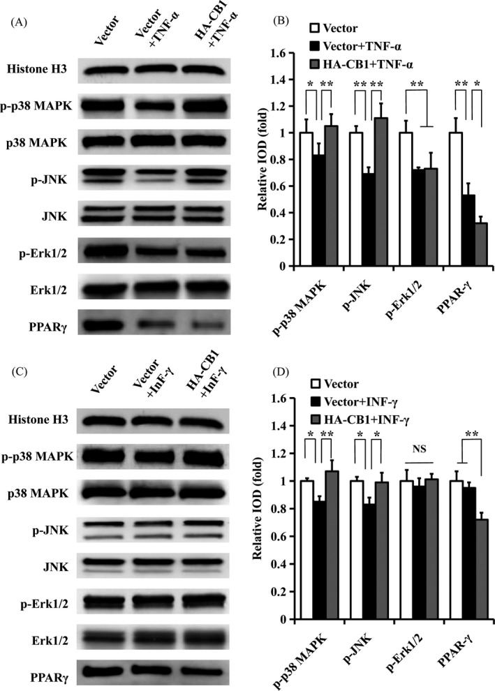
Figure 7.

The effect of CB1 on MAPK signalling pathways and PPAR‐γ in PDLSCs under TNF‐α or INF‐γ stimulation. A, B, 10 ng/mL TNF‐α was used to treat PDLSCs for 4 h. A, Western blot showed the expression of phosphorylated p38 MAPK, JNK and Erk1/2, along with p38 MAPK, JNK, Erk1/2 and PPAR‐γ in PDLSCs. B, The quantitative analysis of phosphorylated p38 MAPK, JNK and Erk1/2 and PPAR‐γ. C, D, 100 ng/mL INF‐γ was used to treat PDLSCs for 4 h. C, Western blot showed the expression of phosphorylated p38 MAPK, JNK and Erk1/2, along with p38 MAPK, JNK, Erk1/2 and PPAR‐γ in PDLSCs. D, The quantitative analysis of phosphorylated p38 MAPK, JNK, and Erk1/2 and PPAR‐γ. Histone H3 was used as an internal control. Error bars represent the SD (n = 3). *P ≤ .05; **P ≤ .01
