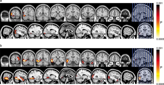
Figure 2.

Cortical regions displaying enhanced activation to emotional compared with neutral words in the P1 (a) and the EPN (b) time windows. These images were thresholded using a voxel‐wise statistical height threshold of (P < 0.001). Functional images are superimposed on a standard brain from a single normal subject (MRIcroN: ch2.nii.gz).
