
Figure 4.
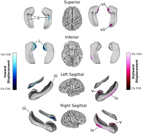
Relationships between right and left whole hippocampal shape and age (with sex, years of education, and APOE ε4 carrier status included in the model). Blue colour maps on the hippocampal surfaces indicate inward displacement after 1–5% FDR correction; red colour maps indicate outward displacement after 1–5% FDR correction. In both hemispheres, inward displacement was localized in (i) the inferior and medial hippocampal head (anterior subiculum and inferior SR/SL/SM of the hippocampal uncus); (ii) the medial hippocampal body (along the CA1‐CA4/DG border); and (iii) the tip of the hippocampal tail (posterior CA1). Outward displacement in both hemispheres was localized to (iv) the lateral hippocampal body (CA1); (v) the lateral edge of the uncus in the hippocampal head (SR/SL/SM); and (vi) the base of the uncus (CA1). (vii) In the right hemisphere only, outward displacements were also observed on the superior tail (a region including parts of the CA4/dentate gyrus, SR/SL/SM, and CA1).