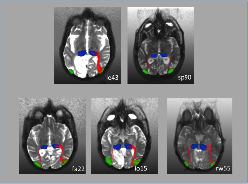
Figure 3.

Fiber tracking results in five representative patients. The hMT ROI was obtained from the fMRI results. The pulvinar ROI was determined by hand based on the individual anatomy. In patient le43 who did not exhibit ipsilesional activation in response to blind field stimulation (see also Fig. 2) fiber tracts could be only tracked in the nonlesioned right hemisphere. This patient had an extensive cortical and subcortical lesion in the left visual cortex (see also Fig. 1). Fiber‐tracts between the pulvinar and ipsilateral hMT were tracked in both hemispheres in patient sp90. This patient, however, did not exhibit hMT activation in the lesioned hemisphere in response to contralateral stimulation. In this patient, the disruption of the pathway occurred probably at the level of the pulvinar, in which we observed a lateral inferior lesion (see also Fig. 1). In patient rw55 area hMT was activated by contralateral motion stimulation in both hemispheres although there was a small lesion in the pulvinar contralateral to the V1 lesion. Fiber‐tracts between the pulvinar and ipsilateral hMT nevertheless were tracked in both hemispheres. This was also the case for the patients fa22 and io15, who did not have any damage to the pulvinar.
